Development of the Body Cavities

Development of the Coelom

By the 13th day of development, the trophoblast has completely invaded the endometrium. The syncytiotrophoblast has eroded into the maternal sinusoids and the primitive hemochorial placenta has been established. The bilaminar embryonic disc is set to begin gastrulation.

The
extraembryonic mesoderm has split into a somatic layer that is attached to the cytotrophoblast and a splanchnic layer that is attached to the secondary yolk sac. These two layers of mesoderm line the space called the chorionic cavity. Since this cavity is lined by mesoderm, it is called a coelom. Since these mesodermal layers line a cavity that is located outside of the embryo proper, the cavity is called the extraembryonic coelom. The extraembryonic coelom is also called the chorionic cavity.

After the formation of mesoderm within the embryo a mesodermally lined cavity will develop within the body of the embryo, the
intraembryonic coelom. Initially, the intraembryonic and extraembryonic coeloms are in communication with one another.

Stacks Image 336

Development of the Thoracic Cavity

  • Intraembryonic Coelom

    A coelom is any cavity that is lined by mesoderm. After the formation of the mesoderm layer during gastrulation, the intraembryonic mesoderm forms three distinct masses. The paraxial mesoderm condenses into blocks of tissue called somites, which align along either side of the neural tube. These will contribute to the formation of musculoskeletal elements of the back and vertebral column.

    Lateral to the paraxial mesoderm is the
    intermediate mesoderm. This mesoderm will form the components of the genitourinary system.

    Lateral to the intermediate mesoderm is the
    lateral plate mesoderm. This mesoderm will split into somatic and splanchnic layers and will form the linings of the body cavities (intraembryonic coelom) and the coverings of the various visceral organs.

    Stacks Image 362
  • Embryonic Folding

    The animation depicts the transverse folding of the embryo and the separation of the intraembryonic coelom from the extra embryonic coelom in the formation of the body cavities.

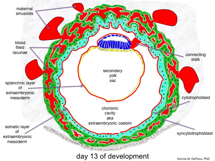
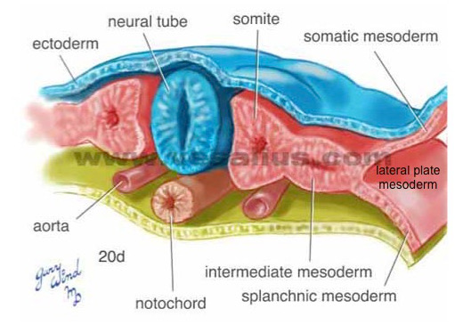
    In this animation note how the lateral plate mesoderm splits to create the intraembryonic coelom which initially is in communication with the extraembryonic coelom. The embryo folds in on itself in the transverse plane and the intraembryonic coelom is ultimately incorporated into the embryo.

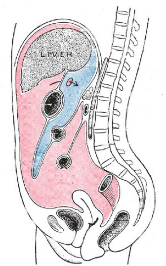
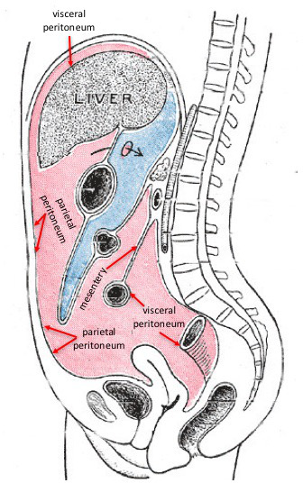
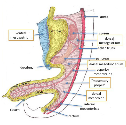
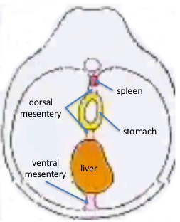
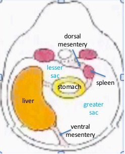
    Note that the gut tube is suspended from the posterior body wall by a sheet of mesoderm which will become the
    dorsal mesentery. Observe that there is a ventral mesentery initially, but it disappears from all but the foregut region of the gut tube.

    The body itself assumes a tubular shape and within we see cross sections of the
    gut tube, the neural tube and the heart tube, all of which will undergo folding as the proceed through development. Even the coelomic cavity is a tubular space extending through the length of the embryo, eventually to be separated into thoracic and abdominopelvic cavities.

  • Septum Transversum

    The septum transversum is a horizontal partition of mesoderm that incompletely separates the contents of the developing thoracic cavity from the contents of the developing abdominal cavity. Note that the foregut, which is connected to the posterior body wall passes through it as does the sinus venosis, which empties into the heart. The septum transversum will make a significant contribution to the development of the respiratory diaphragm. Note that along either side of the septum transversum there is a pericardioperitoneal canal that connects the developing thoracic cavity with the developing peritoneal cavity.

    Stacks Image 384
  • Thoracic Cavity I - Week 5 Early
    Stacks Image 398

    The thoracic cavity is the portion of the intraembryonic coelom that is superior to the septum transversum. Initially, it is a large space containing the heart. Eventually, it will be subdivided into a pair of pleural cavities to contain the lungs and a midline pericardial cavity containing the heart. This partitioning of the thoracic cavity is dependent upon the development, growth and fusion of a pair of membranes that arise from the lateral body wall. These membranes are the pleuropericardial folds. Initially, each fold contains a phrenic nerve and a common cardinal vein. Growth of the lung buds in cranial, caudal and lateral directions causes the pleuropericardial folds to elongate into the pleuropericardial membranes.

  • Thoracic Cavity II - Week 5 Middle

    As the lungs continue to expand into the body wall, they acquire a covering of mesothelium that constitutes the visceral pleura. The mesothelium that is adherent to the body wall constitutes the layer of parietal pleura. The expansion of the lungs also causes elongation of the pleuropericardial folds to form pleuropericardial membranes that begin to approach each other in the midline and which continue to contain common cardinal veins and the phrenic nerves. The pleuropericardial membranes fuse with each other to isolate the heart within the pericardial cavity and the lungs within the pleural cavities.

    Stacks Image 400
  • Thoracic Cavity III - Week 5 Late
    Stacks Image 402

    The pleuropericardial membranes fuse by the end of the 5th week thereby separating the two pleural cavities and the pericardial cavity from one another. As a result, the phrenic nerves course between the fibrous pericardium, and the mediastinal pleura.
    Each lung is covered by a layer of
    visceral pleura and parietal pleura lines each pleural cavity. The common cardinal veins fuse to form the superior vena cava. Caudally, each pleural cavity is continuous with the peritoneal cavity in the abdomen through the pericardioperitoneal canals.

Development of the Peritoneal Cavity

  • Overview

    The abdominopelvic cavity is the space inferior to the diaphragm. The upper part of the cavity is lined by mesothelium called the peritoneum. Parietal peritoneum contacts the body wall and visceral peritoneum invests most of the abdominal organs.
    The
    liver is surrounded by a capsule formed by the visceral peritoneum. Visceral peritoneum is also attached to the wall of most of the GI tract. Double folds of peritoneum, called mesenteries anchor the abdominal visceral organs to the abdominal body wall.
    The
    parietal peritoneum lines the entire inside of the abdominal cavity.

    Roll over the image

    Stacks Image 477
    Stacks Image 480

    Sagittal section through the abdominopelvic cavity of an adult showing the locations of the parietal and visceral peritoneal layers

  • Dorsal & Ventral Mesenteries

    Mesenteries are double folds of peritoneum that attach the visceral organs to the body wall. There is a dorsal mesentery that initially anchors the developing gut tube to the posterior abdominal wall and a ventral mesentery that anchors the gut tube to the anterior body wall. The ventral mesentery disappears from the midgut and hindgut regions of the tube, but persists in the foregut region as the ventral mesogstrium.
    The
    dorsal mesentery is named for the regions of the gut tube with which it is associated. Dorsal mesogastrium is associated with the developing stomach, dorsal mesoduodenum is associated with the developing duodenum and dorsal mesocolon with the developing colon to name a few. Some parts of the dorsal mesentery will disappear as development progresses (dorsal mesoduodenum, e.g.) while other parts may become enhanced (dorsal mesogastrium to form the greater omentum e.g.).

    Stacks Image 431
  • Divisions of the Peritoneal Cavity

    Initially, the gut tube is suspended in the midline from the posterior body wall. Following rotation of the stomach, the liver is pushed to the right side of the abdomen and the stomach is pushed to the left. As a result, a small portion of the peritoneal cavity is located behind the stomach. It is called the lesser sac or omental bursa. The larger space within the peritoneal cavity, anterior to the stomach and liver, is called the greater sac.
    Knowledge of these subdivisions is important in pathologies of organs that are located behind the stomach in the omental bursa. in particular, the pancreas, duodenum and aorta.

    Stacks Image 443

    Before rotation of the stomach

    in the sagittal view of the adult abdomen to the right, the lesser sac is colored blue and the greater sac is colored pink. This subdivision of the peritoneal cavity is the result of the rotational events that occur during development of the GI tract. The arrow is passing through the epiploic foramen (of Winslow), the only natural connection between the greater and lesser sacs.

    Stacks Image 445

    After rotation of the stomach

    Stacks Image 452
    Stacks Image 454

    Sagittal section through the abdominopelvic cavity of an adult showing the subdivisions of the peritoneal sac