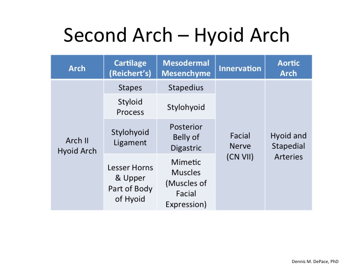
The second branchial arch is known as the hyoid arch because it gives rise to parts of the hyoid bone and muscles that attach to it.
Cartilage
The cartilage of the second arch, which is derived from neural crest, is known as Reichert's cartilage. It gives rise to the following structures:
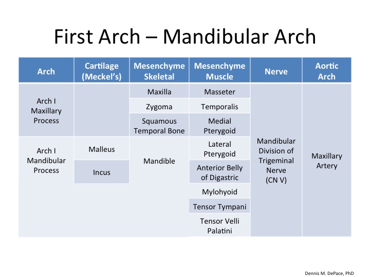
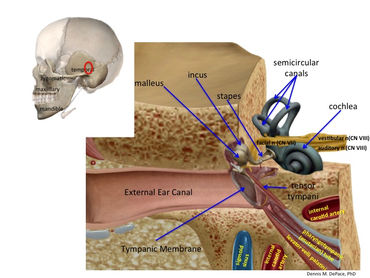
- stapes of the middle ear cavity (note that malleus and incus are from Arch I)
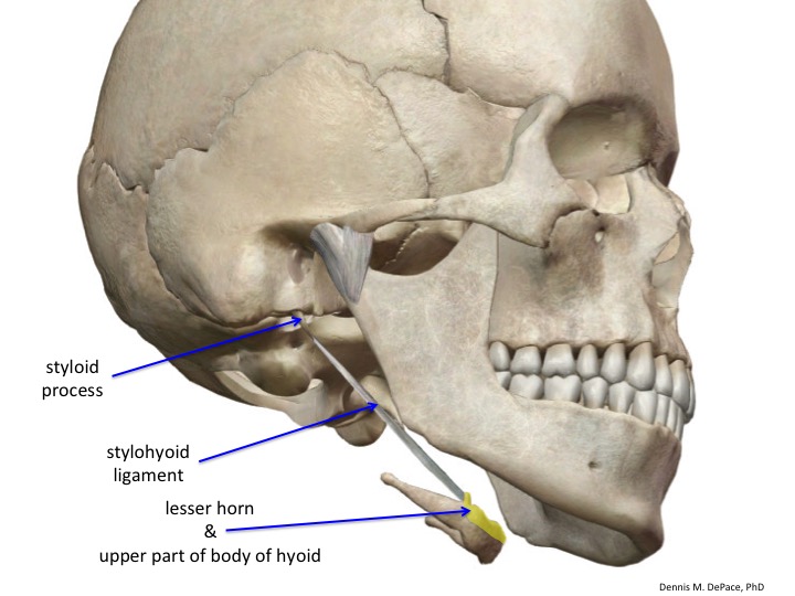
- styloid process
- stylohyoid ligament
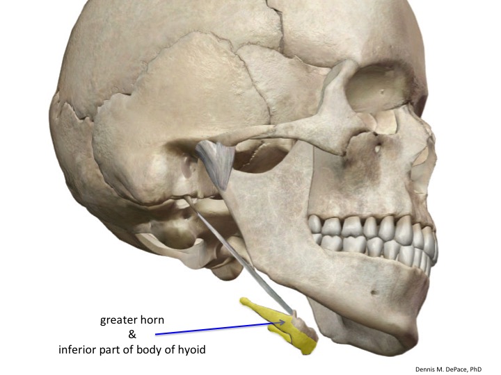
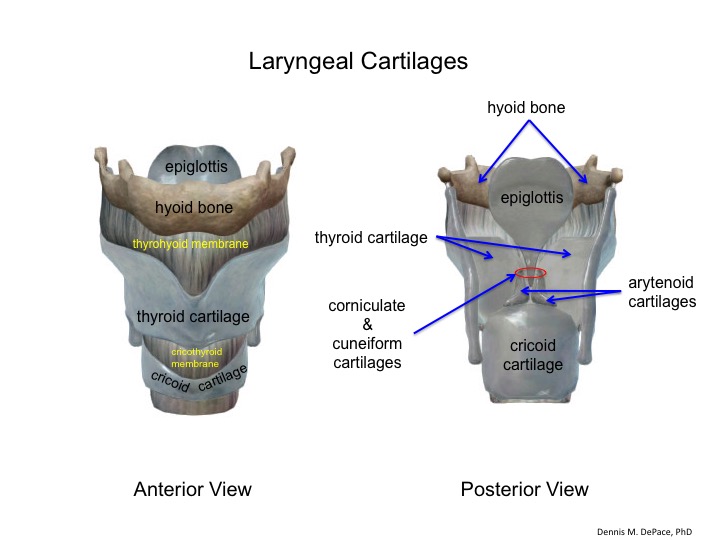
- lesser horn of hyoid bone
- upper part of the body of the hyoid bone
Mesenchyme
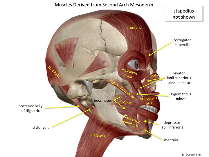
Several pairs of muscles are derived from the mesenchyme of the second branchial arch. These include:
- stapedius
- stylohyoid
- posterior belly of digastric
- mimetic muscles, including
- frontalis
- orbicularis oris
- orbicularis oculi
- buccinator
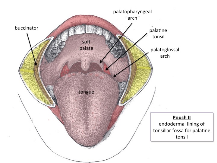
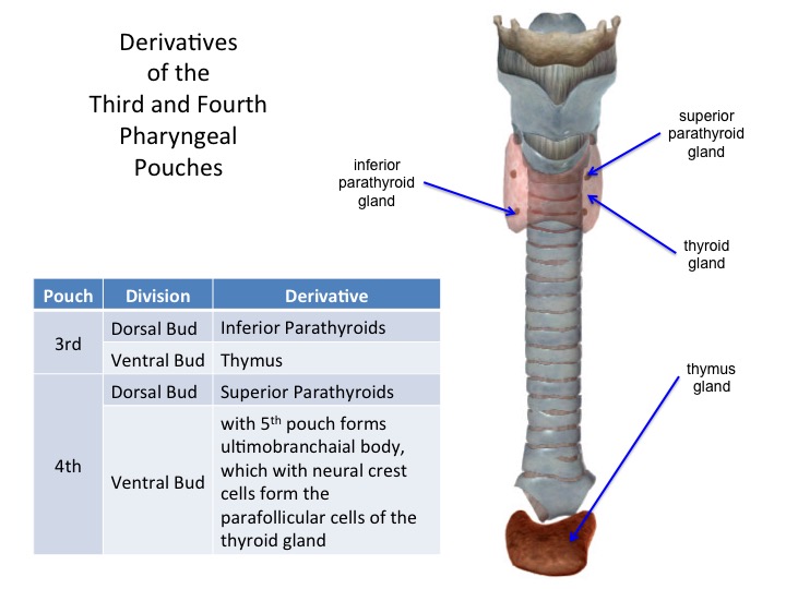
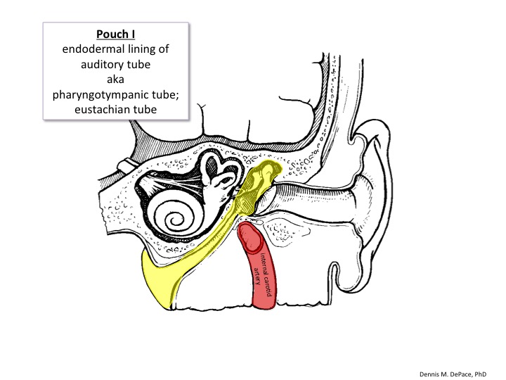
Pouch Endoderm
The endoderm of the second pharyngeal pouch forms the lining of the tonsillar fossa for the palatine tonsils.