All muscle tissue in the body is derived from mesoderm, with the exception of a small population of smooth muscle cells in the eye. These arise from ectoderm.
Muscle tissue is divided into three types, studies in Microanatomy.

  • Smooth Muscle - forming the wall of most visceral organs and blood vessels and located in a few other tissues such as the skin and eye.
  • Cardiac Muscle - located within the chambers of the heart.
  • Skeletal Muscle - voluntary muscle attached to the bones of the body and which produce our ability to move.

We will only consider skeletal muscle in this unit since it it related to the axial and appendicular skeletons.

Smooth muscle will be discussed in development of some of the hollow viscera and cardiac muscle will be discussed in the context of development of the cardiovascular system.

In order to add confusion to the process, embryologists have now come up with new designations for the origins of muscles.

  • The primaxial domain refers to mesoderm derived exclusively from the somite mesoderm, the myotome. Muscles derived from the primaxial domain include the epaxial muscles or erector spinae and related deep back muscles innervated by the dorsal primary rami of spinal nerve and also some of the muscles of the shoulder girdle and intercostal muscles, innervated by ventral primary rami of spinal nerves.

  • The abaxial domain refers to mesoderm of both somite and lateral plate origin, forming the infrahyoid muscles, abdominal wall muscles and the limb muscles. These muscles are all innervated by the ventral primary rami of spinal nerves.

  • Myotome - Epimere vs Hypomere

    Skeletal muscles arise from the mesoderm layer of the embryo. Some of that mesoderm is from neural crest origin in the branchial arches and will give rise to the muscles in the head and neck, to be studied later.
    Muscles of the torso and appendages are derived from either the myotome portion of the somite or the lateral plate mesoderm.
    Under the influence of Wnt proteins from the dorsal part of the neural tube, the dorsomedial portion of the myotome gives rise to the epimere, which will develop the muscles of the deep back, the erector spinae.
    Under the influence of Wnt proteins from the surface ectoderm and BMP4 from the lateral plate mesoderm the ventrolateral portion of the myotome gives rise to the hypomere, which develops into the muscles of the torso, such as the intercostals and abdominal musculature and which contributes to the musculature of the upper and lower limbs. The lateral plate mesoderm also contributes to the musculature of the upper and lower limbs.

    Stacks Image 36
  • Innervation - Epimere vs Hypomere
    Stacks Image 43

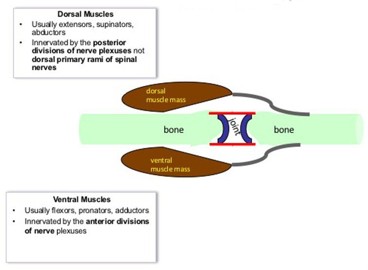
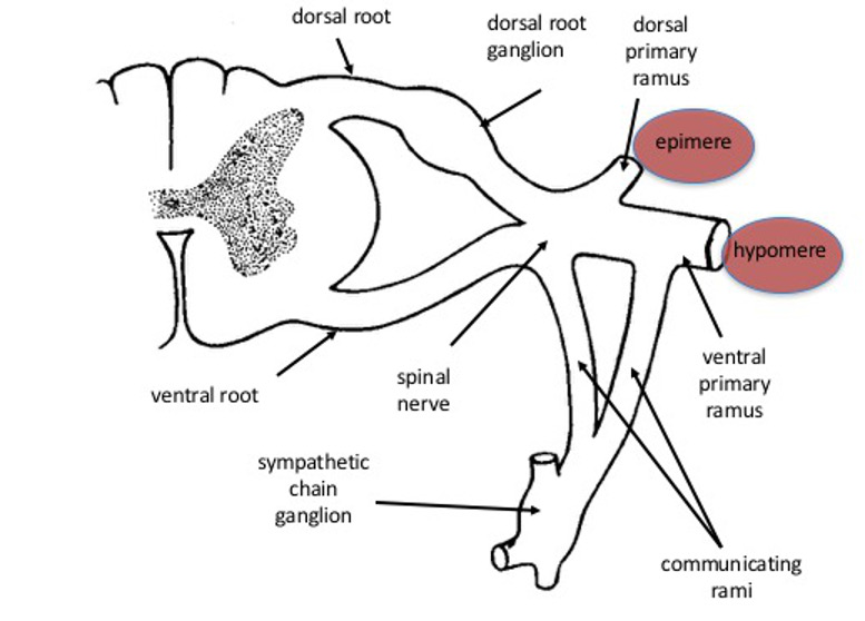
    The diagram of a typical spinal nerve shows that the muscles of the epimere, i.e. the erector spinae and deep muscles of the back are innervated by the dorsal primary rami of spinal nerves. These muscles are also identified as originating from the primaxial domain.
    The ventral rami of the spinal nerves innervate the muscles derived from the hypomere. In the upper and lower limbs, the ventral primary rami from the brachial plexus and lumbosacral plexus for the innervation of the upper limb and lower limb respectively. The ventral primary rami also form the intercostal nerves, that innervated the intercostal muscles and abdominal muscles. These muscles are also identified as originating form the abaxial domain.

  • Organization of the Limb Bud

    In the cross section through an embryo between 5 and 6 weeks of development, the upper limb buds demonstrate some of the important features associated with limb development. The thickened surface ectoderm has formed the apical ectodermal ridges (AER).
    In the midline of the limb buds, skeletal elements are beginning to develop. Differentiation of the limb proceeds in a proximal to distal direction. Mesoderm that migrates into the limb to form the muscles aggregates into two masses. One mass is located dorsally in the limb and the other is located ventrally. This mesoderm will begin to differentiate into myoblasts which will fuse with one another to forms multinucleate skeletal muscle cells. Eventually the muscles will associate with the skeletal elements to produce functional groups of muscles. The posterior divisions of the nerve plexuses will innervate the muscles derived form the dorsal muscle mass and the anterior divisions of the plexuses will innervate the ventral muscles.

    Stacks Image 50
  • Muscle Groups in the LImb

    The dorsal muscle mass will give rise to the posterior muscles of the limb, In the upper limb these are generally the extensors supinators and abductors. These are generally supplied by the branches of the posterior divisions of the brachial plexus, including the axillary and radial nerves.
    The ventral muscle mass will give rise to the anterior muscles of the limb. In the upper limb these are generally the flexors, pronators and adductors. These muscles are supplied by the branches of the anterior divisions of the brachial plexus that form the medial and lateral cords and include the musculocutaneous, ulnar and median nerves.

    Stacks Image 57