Development of the Eye

Stacks Image 708
The eye begins development as the optic groove in the diencephalon at about day 22. These expand into the optic vesicles at about day 28. The optic vesicles have contact with the surface ectoderm. The optic vesicles invaginate to become the optic cups. They also induce the surface ectoderm to form the lens placode. By day 30, the lens vesicle is formed. The optic cup is connected to the diencephalon as by way of the connecting stalk. Blood vessels pass ventrally into the optic stalk to nourish the developing eye.
Once the optic cup is formed as an outgrowth of the diencephalon, it induces the formation, in succession, of the lens and cornea. The surrounding mesoderm also contributes to the overall structure of the eye.
  • Day 31

    Around day 30, the optic vesicle begins to invaginate to form the optic cup. At the same time, induction of the lens placode begins by a thickening of the surface ectoderm.

    By day 31, the optic cup forms two layers. the
    outer layer of the optic cup will form the pigment layer of the retina, while the inner layer of the optic cup will form the neural retina. At this time, the lens placed begins to invaginate to forms a lens vesicle.

    Stacks Image 769
  • Day 32
    Stacks Image 771

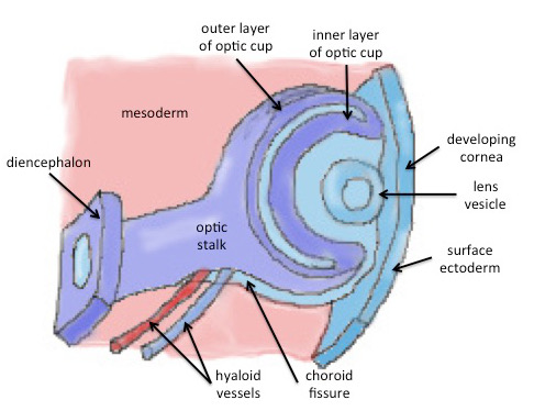
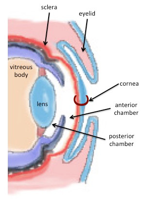
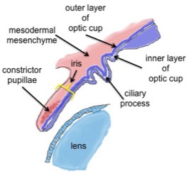
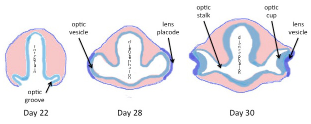
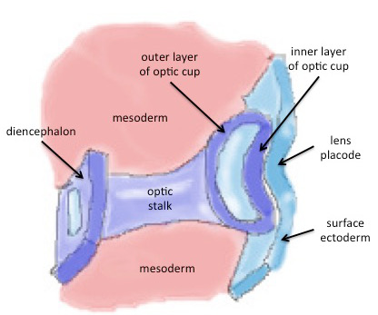
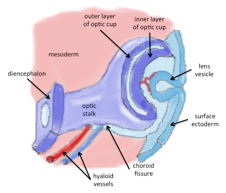
    At around day 32, the optic cup is divided into inner and outer layers. Note also that the lens has become vesicular and is sinking beneath the surface ectoderm. The hyaloid vessels approach the developing eye by entering the optic cup through the choroid fissure.

  • Day 33

    Later in development, the inner and outer layers of the optic cup come into close apposition. The hollow lens vesicle has separated from the surface ectoderm and the ectoderm over the lens area will begin to differentiate into the cornea.

    Stacks Image 773
  • Newborn Eye
    Stacks Image 775

    In the newborn the eye is completely formed. Note the embryonic tissues that form each portion of the eye

  • Chart of Derivitives of the Eye
    Stacks Image 777

Optic Nerve

Stacks Image 790
The optic vesicle is connected to the developing diencephalon by way of an optic stalk.
On the ventral surface of the optic stalk there is a groove, the
choroid fissure.
Hyaloid vessels
pass into the choroid fissure to reach the developing retina. They will become enclosed within the optic nerve when the choroid fissure closed in week 7 of development. They are thereafter called the central retinal vessels, which enter the eye in the region of the optic disc.
Optic nerve fibers,
originating in the ganglion cells of the retina, travel back to the CNS in the inner layer of the optic stalk. This layer is continuous with the pia arachnoid surrounding the brain.
The outer layer of the optic stalk is continuous with the
sclera of the eye and becomes the dura mater covering the optic nerve.

Cornea

Stacks Image 798
The cornea is the anterior, transparent surface of the eye that is continuous posteriorly with the sclera.

Three layers form it
  • Layer of surface ectoderm
  • Substantia propria – continuous with the sclera
  • Inner layer of epithelium

Anterior chamber is fluid filled space between the cornea and the anterior surface of the iris.
Posterior chamber is between the iris and the lens.

Both spaces are filled with aqueous humor.

Iris and Ciliary Body

Stacks Image 805
The iris develops from both layers of the optic cup. The outer layer of optic cup forms the pigmented layer of the iris. The inner layer of optic cup forms the non-pigmented layer of the iris.

Both contribute to the pupillary smooth muscles

Mesenchyme of
neural crest origin forms the anterior surface of the iris and is continuous with the sclera

Smooth muscle of the iris forms from the underlying ectoderm. This ectoderm former the dilator and constrictor papillae muscles.