Appendicular Skeleton

The appendicular skeleton is the skeleton of the upper and lower limbs.

  • In the upper limb, the bones include: clavicle, scapula, humerus, radius and ulna, 8 carpals, 5 metacarpals and 16 phalanges.
  • In the lower limb, the bones include: hip bone, femur, tibia and fibula, 7 tarsals, 5 metatarsals and 16 phalanges.
These bones arise from the mesoderm of the limbs and except for the clavicle, which develops by intramenbranous ossification, they develop by endochondral ossification.

Limbs

  • Overview
    Stacks Image 85

    Limb buds make their appearance in the fourth week of development. The upper limb bud appears in the cervical region around day 24, followed by the lower limb bud appearing in the lumbar region around day 28. By 5 weeks they are distinct features of the embryo.
    Developmentally, the upper limb is about 2 to 4 days ahead of the lower limb. Each limb is induced by the presence of surface ectoderm that is known as the
    apical ectodermal ridge (AER) . The AER guides the development of the limbs by suppressing differentiation of the underlying mesoderm. As the limb elongates, the AER moves further from the proximal parts of the limb which then begin to differentiate into muscle and bone.

  • Orientation of the LImb Buds

    Note that initially, each limb projects from the torso in a straight line. The cranial surface of each limb is designated the preaxial border. On the upper limb the thumb is on the preaxial border and on the lower limb, the great toe is on the preaxial border. The caudal surface of each limb is designated the postaxial border. The little finger and little toe would be on the postaxial side of the limb.

    Stacks Image 83
  • Apical Ectodermal Ridge

    The apical ectodermal ridge (AER) secretes factors that prevent the underlying mesenchyme from differentiating into limb structures. As the limb elongates, the AER becomes displaced distally and its influence on the proximal parts of the limbs decreases. Differentiation of the limbs proceeds in a proximal to distal gradient. The hand and foot take on "paddle" shapes and eventually the AER splits into 5 segments in each limb to guide the development of the fingers and toes. If it divides into more than 5 parts, polydactyly (extra digits) can result.

    Stacks Image 87
  • Differentiation of the Limb Structures
    Stacks Image 127

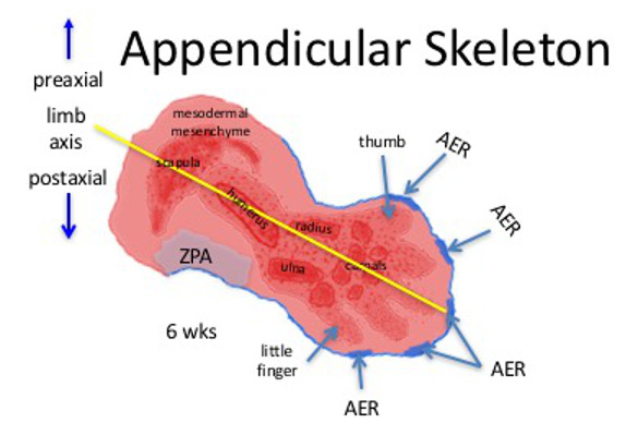
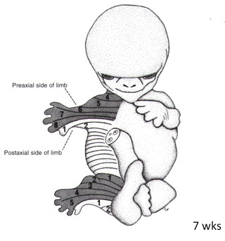
    Development of the limb is guided by the apical ectodermal ridge (AER), which inhibits the differentiation of the mesoderm directly below it. In this way, development of the limb proceeds in a proximal to distal direction. In the diagram of an upper limb of a six-week embryo (above), the proximal mesenchyme is no longer under the inhibitory influence of the apical ectodermal ridge (AER). Note that the scapula, humerus, radius, ulna and carpal bones have begun to form as cartilage models from the condensing mesenchyme. By the 7th week of development they will begin to undergo endochondral ossification.
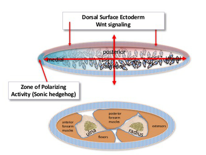
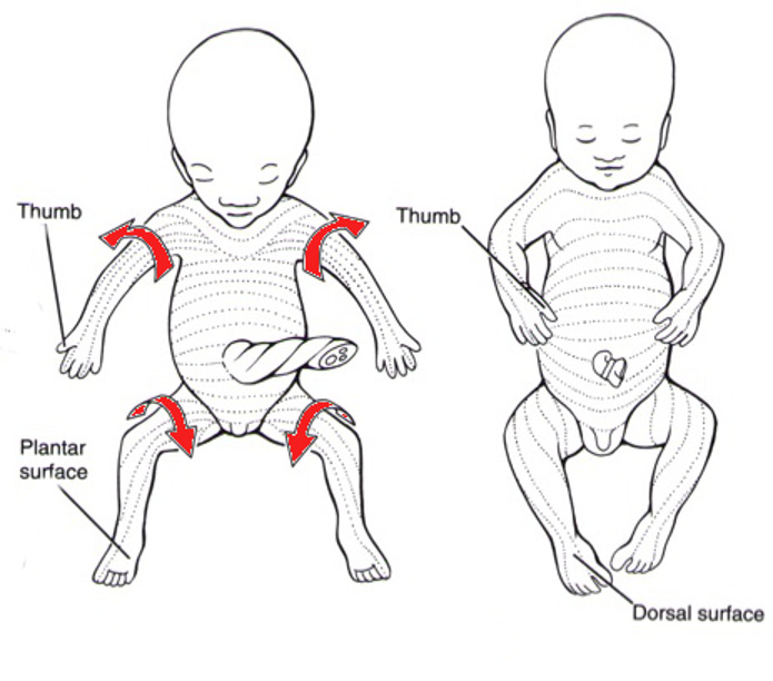
    Note that the
    radius is on the pre-axial side of the limb and the ulna is on the post-axial side of the limb. Sonic hedgehog (Shh) from the zone of polarizing activity (ZPA) determines the formation of post-axial vs pre-axial structures. The post axial limb structures in the upper limb form earliest and include the humerus, ulna, and digits 2 through 5 and their related carpals and metacarpals.
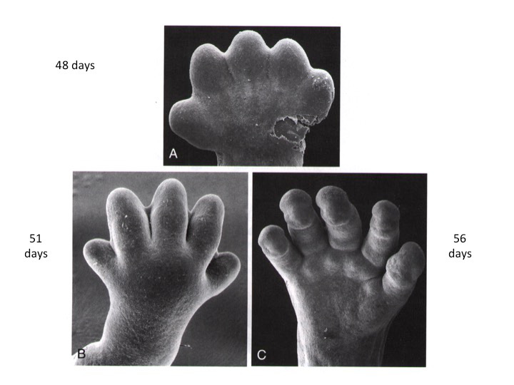
    The
    apical ectodermal ridge (AER) has split into 5 pieces to guide the development of each of the digits. Observe that the thumb is on the pre-axial side of the paddle shaped hand and the little finger is on the post-axial side. The phalanges remain undifferentiated at this stage.
    Splitting of the apical ectodermal ridge into more than 5 segments is related to the appearance of extra digits,
    syndactyly.

    Stacks Image 135

    This diagram illustrates how the posterior limb ectoderm and the zone of polarizing activity (ZPA), produce gradients of two different morphogens that will signal the limb mesoderm on how to proceed with development. Cells at different distances from the sources of these morphogens will form different parts of the limb, posterior vs anterior and medial vs lateral. Wnt signaling in the posterior surface ectoderm produces transcription factors to guide the mesenchyme to form posterior limb structures, while the zone of polarizing activity simultaneously creates a gradient of Sonic hedgehog to promote the development of postaxial structures in the limb bud.
    The end result is seen the cross section of the forearm below.

  • Digits
    Stacks Image 89

    Initially, each hand and foot is "paddle" shaped. As the AER degenrerates between digits, it is split into 5 pieces, thus guiding the elongation of the digits into distinct entities. As the limb nears completion of development, separation of the digits is accomplished by degeneration of the tissue between the digital rays through the process of apoptosis. Failure of this to occur results in the fusion of digits, a condition known as syndactyly.

  • Dermatomes
    Stacks Image 93

    The numbers indicate the dermatome and spinal segments that are represented in each limb. C4 to T2 dermatomes are represented in the upper limb and the corresponding spinal nerves innervate the musculature, while L1 to S2 dermatomes are represented in the lower limb and their spinal nerves innervate the musculature.

    Stacks Image 91
  • Rotation of the LImbs
    Stacks Image 102

    Final positioning of the limbs is the result of rotation of each limb, in different directions. The upper limb undergoes a slight degree of lateral rotation so that the thumb points laterally and the original ventral surface (i.e. flexor surface) of the limb still faces anteriorly. The lower limb undergoes a great degree of medial rotation, however.
    As a result of this rotation, the great toe moves medially and the original ventral surface (i.e. flexor surface) faces posteriorly. Hence, the dermatomes of the lower limb seem to “spiral” down the limbs.

  • Joints

    Joints form as a result of breakdown of tissue between the cartilaginous models of the bones. The space that is generated will become the synovial cavity and the cartilage over the articular ends of the bones will forego ossification to remain as smooth articular cartilage.
    The mesenchyme surrounding the synovial cavity will develop into dense connective tissue to form the joint capsule and ligaments.

    Stacks Image 114