Axial Skeleton

Stacks Image 17
The axial skeleton consists of the:
  • vertebral column
  • ribs
  • sternum
  • skull
  • hyoid bone

It is derived from both
paraxial (somitic) mesoderm and neural crest sources.

The
neural crest is by way of the branchial arches and it contributes to the development of the anterior parts of the skull and hyoid bone while the paraxial mesoderm forms masses of tissue called somites that contribute to the posterior parts of the skull and the vertebral column, ribs, costal cartilages and sternum.

Vertebral Column

  • Mesoderm: Major Divisions
    Stacks Image 101
    Stacks Image 181

    By the end of the fourth week of development, the mesoderm that formed during gastrulation, occupies a position between the ectoderm and endoderm of the trilaminar embryonic disk. It is subdivided into several masses.

    • Notochord forming the midline axis of the embryonic disc.
    • Paraxial mesoderm forms distinct blocks of mesoderm called somites which will from the elements of the back and vertebral column.
    • Intermediate mesoderm, located lateral to the somites, will contribute to the formation of genitourinary structures.
    • Lateral plate mesoderm splits into two layers. One layer is affixed to the surface ectoderm and is called the somatic layer of lateral plate mesoderm. This layer of mesoderm plays a role in the development of the limb skeleton and musculature.

  • Somites

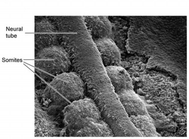
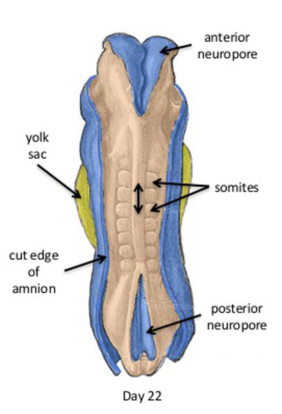
    In this day 22 embryo, note the pairs of somites along the neural tube. Recall that the neural tube forms by fusion of the neural folds, beginning in the center of the embryo and proceeding simultaneously in a rostral and caudal direction.
    Here at day 22, the neural folds remain unfused both cranially and caudally, forming the
    anterior and posterior neuropores
    Somites first appear in the cranial region around day 20. Eventually 44 pairs of somites will develop along the sides of the neural tube by day 30.
    This cranial to caudal, segmental progression is regulated by
    HOX genes which also determine where the upper and lower limb buds will appear.

    Stacks Image 108
    Stacks Image 115

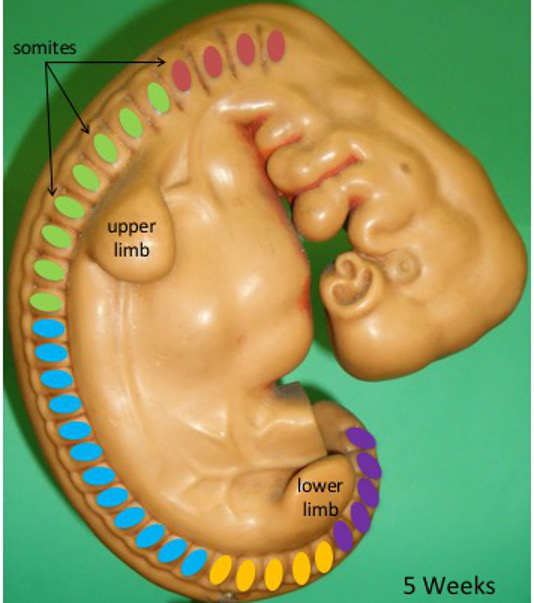
    By day 30, roughly 44 pairs of somites can be seen through the surface ectoderm of the embryo.
    They are grouped into:

    • 4 occipital pairs (pink)
    • 8 cervical pairs (green)
    • 12 thoracic pairs (blue)
    • 5 lumbar pairs (yellow)
    • 5 sacral pairs (purple)
    • 3-5 coccygeal pairs (not indicated)

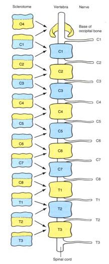
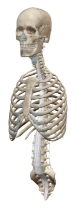
    Note that these nearly correspond to the number of vertebrae in the vertebral column.

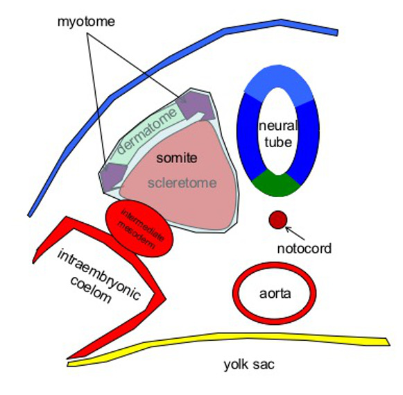
    Each somite is divided into sclerotome, dermatome and myotome portions.
    The
    vertebrae form from the sclerotome portion of the somite through the inductive influences of Sonic hedgehog (Shh) secreted by the notochord and ventral neural tube.
    The cells express PAX 1 a factor that controls chondrogenesis and subsequent bone formation.
    Sclerotome cells migrate around the neural tube to form the vertebral arch and around the notochord to form the vertebral bodies.

    Stacks Image 124
  • Re-Segmentation of the Somites

    The emerging spinal nerves course in the rostral part of the sclerotome. Each sclerotome splits to cranial and caudal segments at the point where the spinal nerve passes.
    Subsequently, the cranial part of one sclerotome fuses with the caudal part of the sclerotome in front of it to form the vertebral bodies. By default, the intervertebral foraminae are then located between the contiguous vertebrae.

    This animation illustrates the process. Note the splitting of the sclerotomes at the point where the spinal nerves pass and the cranial and caudal segments of two different sclerotomes fusing to form the vertebral body.
    Note also that the notochord is incorporated into the bone of the vertebral body but persists as the nucleus pulposus of the intervertebral discs.

    In this image we note that the caudal segment of the 4th occipital sclerotome (O4) fuses with the cranial part of the first cervical sclerotome (C1) to form the base of the occipital bone in the skull.

    As a result the C1 spinal nerve emerges through the space between the occipital bone and C1. Similarly,
    all the cervical nerves emerge from the intervertebral foramen superior to their correspondingly numbered vertebra ending with the C8 nerve.

    Beginning with the
    first thoracic nerve, all spinal nerves emerge from the intervertebral foramen inferior to their correspondingly numbered vertebra.

    Stacks Image 139
  • Cauda Equina
    Stacks Image 146

    This slide shows the progressive stages in the formation of the cauda equina. Initially, the spinal cord and vertebral column are equal in length. As the fetus grows, the vertebral column increases in length, while the spinal cord remains relatively stable. The caudal end of the spinal cord, the conus medullaris, shifts upward in the vertebral canal causing elongation of the lower nerve roots to form the cauda equina. At birth, the conus medullaris is opposite the L3 vertebral level and by adulthood it has shifted upward to the level of the disc between L1 and L2.


Ribs and Sternum

Cells from the lateral part of the sclerotome, migrate into the lateral plate mesoderm and form primordial of ribs and the sternum. Extra ribs are occasionally found in the cervical region, attached to the C7 vertebra.


Skull

  • Overview

    The skull can be divided into two parts:
    Viscerocranium - skeleton of the face
    Neurocranium - portion of the skull forming the cranial vault

  • Viscerocranium

    The viscerocranium consists of the bones that form the skeleton of the face and lower jaw and hyoid. They are derived from the first three branchial arches.

    The first branchial arch divides into a
    maxillary process and a mandibular process.

    The mesenchyme of the maxillary process gives rise to the maxilla, zygomatic and the squamous part of the temporal bone. This mesenchyme is derived from the neural crest and the bones form via intramembranous ossification.

    The
    mandibular process contains Meckel's Cartilage. However, as development continues, neural crest derived mesenchyme around the cartilage undergoes intramembranous ossification to form the mandible. The cartilage largely regresses except for what forms the sphenomandibular ligament along with the malleus and incus and parts of the middle ear cavity.

    The second branchial arch is called the
    hyoid arch. The cartilage of the 2nd arch (Reichert's cartilage) forms the stapes and styloid process along with lesser horns and upper body of the hyoid bone. These bones thus develop via endochondral ossification.

    The remainder of the hyoid bone is formed via endochondral ossification of the cartilage of the third branchial arch.

    The cartilages of the 4th-6th arch will not undergo ossification and instead will fuse to form the laryngeal cartilages.

    Stacks Image 49
  • Neurocranium - Cartilagenous (Chondrocranium)

    The neurocranium is the part of the skull that surrounds and protects the brain. The base of the neurocranium consists of bones that form by endochondral ossification and which are derived from mesenchyme of both neural crest and somitic origin. This part of the neurocranium is called the chondrocranium.

    Those of
    neural crest origin include the ethmoid bones, greater and lesser wings of the sphenoid bone and the anterior part of the sella turcica.

    Those derived from
    somatic origin, originate in the sclerotomes of occipital somites and include the posterior part of the sella turcica, body of the sphenoid, base of the occipital bone and petrous part of the temporal bone.

    The chondrocranium includes numerous centers of ossification. Cartilagenous Joints in the cranial base, such as the sphenoid-occipital synchondrosis, are responsible for continued antero-posterior growth of the neurocranium.

    Stacks Image 51
  • Neurocranium - Membranous (Dermatocranium)
    Stacks Image 53

    The membranous neurocranium (or dermatocranium) consists of the flat bones of the roof of the skull that form by intramembranous ossification. Those located more anteriorly in the skull such as the frontal bone are from neural crest origin, while those more posterior such as the parietal bones and upper part of the occipital bone are from somitic mesoderm.

  • Fontanelles
    Stacks Image 65
    At birth, the flat bones of the skull are separated by connective tissue, which will eventually form the sutures of the skull. The fontanelles are located where more than two bones meet and are generally wide. The fact that the sutures are not completely ossified at birth, allows the bones of the skull to overlap during birth. After birth, the bones move back to their original position.

    The fontanelles also allow the brain to continue to grow at the same rapid pace as it had prenatally. Even once the fontanelles close, the sutures between the bones allow for rapid growth during the first few years of life (through ~age 5). The emphasis on growth of the neurocranium is reflected in its disproportionate size in the neonate (~4/5ths of skull volume) relative to the smaller size of the face (~1/5th).
    Stacks Image 68
    The anterior fontanelle is the largest and is often referred to in lay terms as the “soft spot” on the baby’s head. Palpation of the anterior fontanelle can provide information about the normal progress of ossification of the skull and whether intracranial pressure is normal. The posterior fontanelle generally closes around the second month after birth while the larger anterior fontanelle closes around the 18th month post partum.

    At around 5 to 7 years old, the child has nearly all of the cranial volume and subsequent growth of the skull is due to increase in the size of the facial skeleton. In adults, the facial skeleton and the cranial vault are each about 50% of the skull volume.